Retinal complications of Diabetes

Diabetes is best defined as a metabolic cum vascular syndrome associated with multiple causative factors characterized by chronic hyperglycaemia with disturbances of carbohydrate, fat and protein metabolism resulting from defects in insulin secretion, insulin action or both, leading to changes in both small blood vessels and large blood vessels. This is often associated with long term damage, leading to malfunction and failure of various organs like eyes, kidneys, heart, nerves and blood vessels.
When the elevated blood sugars effect eyes, it is called diabetic retinopathy. Here we commonly see retinal damage due to uncontrolled blood sugar. Retina is a layer of tissue in the back of eyeball. It contains photoreceptor cells called rods and cones. Rod receptors are located throughout the retina and provides black-white vision and function well in low light. Cone receptors are concentrated in small central area of the retina. These are responsible for central vision & Color vision, function well in medium and bright light.
The function of retina is to capture the photons and convert into chemical and nervous signals which are transported to visual centers of brain through optic nerve. These signal are converted into images and visual perception in the visual cortex of the brain. Maximum of the visual activity is done in the part of retina.
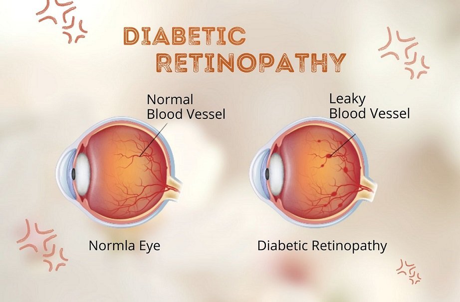
In diabetic retinopathy patients the blood vessels of the retina may change or swell and leak fluid. Damage of these blood vessels of retina will lead to significant vision loss.
Stages of diabetic retinopathy
- Mild non-proliferative retinopathy - it is beginning stage of retinopathy, swelling starts in the tiny areas of blood vessels of retina.
- Moderate non-proliferative retinopathy - increased swelling of tiny blood vessels of retina.
- Severe non-proliferative retinopathy - accumulation of blood & other fluid and causes decreased blood flow & nourishment to retina.
- Proliferative retinopathy - it is advanced stage of retinopathy; here new blood vessels start growing & there will be a high risk of fluid leakage.
Naturopathy introduces Iris diagnosis as diagnostic and prognostic method of evaluation in patients to see the changes in iris by using magnifying lens, flashlights, slit lamps and microscopes. Iris is a protected internal organ of the eye, located behind the cornea. Clock wise, both right and left iris represents a complete human body structure. Any changes from normal iris is an indication of abnormal function of any part or organ of human body. There may be changes like lines, flakes, open or closed lacunae, block dots etc.
Pancreatic triad appears in the iris as large lacunae and honeycomb crypt in one or more of the pancreas representing area. This indicates a potential for deficiency in the pancreas.
Common problems of diabetic retinopathy are blurred vision, fluctuating vision, blind spots, seeing floating spots, double vision, changes in color perception, sudden loss of vision & eye pain in advanced cases.
Ayurvedic Treatment for Diabetic Retinopathy

Ayurveda talks about diabetic retinopathy as Dristipatalagata roga. Diabetic Retinopathy can be compared to Timira involving all the four patalas. Patalas are described on the basis of functional composition of dhatus of dristi. The symptoms of vision are manifested when the vitiated dosha afflicting the concerned dhatu in dristi patalas. Based on the diagnosis treatments such as Tarpana or Pariseka.


